Pelvic Anatomy Female Ligaments : Ligaments as a Source of Pain and Suppressed Performance ... - The female bony pelvis is divided into:. Of female pelvic organ sacrospinous ligament just medial to the ischial spine, exiting the pelvis through the greater sciatic foramen. Ligaments of the reproductive system. From internal to external lateral to the uterus and close to the lateral pelvic wall. Ligaments and anatomy important in pelvic. Start studying female pelvic anatomy.
Lotze, md facog female pelvic medicine & reconstructive surgery division & fellowship director, women's pelvic health & continence center obturator membrane. Dummies has always stood for taking on complex concepts and making them easy to understand. This short article describes the the cardinal ligament likely provides support to the pelvic viscera, as structural abnormalities. The female bony pelvis is divided into: Above the pelvic brim and has no obstetric importance.
BIO202-Female Reproductive | Anatomy models labeled ... from i.pinimg.com The female bony pelvis is divided into: Functional anatomy of the male pelvic floor online course: Choosing a primary surgical procedure. Sagittal section female pelvis with peritoneum. The pelvis (plural pelves or pelvises) is either the lower part of the trunk of the human body1 between the the female pelvis is larger and broader than the male pelvis which is taller, narrower, and the lateral lumbosacral ligament, partly continuous with the iliolumbar ligament, passes down from. Anatomy of the female pelvic region. The posterior sacroiliac ligament supports the sacroiliac joint. This anatomy section promotes the use of the terminologia anatomica, the international standard of anatomical nomenclature.
Anatomic features that are clinically applicable to female pelvic surgery are.
This short article describes the the cardinal ligament likely provides support to the pelvic viscera, as structural abnormalities. The fallopian tubes are made up of three layers. Dummies helps everyone be more knowledgeable and confident in applying what they know. Double fold of peritoneum extending laterally from the uterus towards the pelvic side wall and encloses the uterine tube. Surgical management of stress urinary incontinence in women: Four ligaments inguinal ligament • important for repair of inguial hernia cooper's ligament • frequently used in bladder suspension. The pelvis (plural pelves or pelvises) is either the lower part of the trunk of the human body1 between the the female pelvis is larger and broader than the male pelvis which is taller, narrower, and the lateral lumbosacral ligament, partly continuous with the iliolumbar ligament, passes down from. The pelvis's frame is made up of the bones of the pelvis, which connect the axial skeleton to the femurs, and therefore acts in weight bearing of the upper body. Peritoneum and the broad ligament. Whether it's to pass that big test, qualify for that big promotion or even master that cooking technique; Above the pelvic brim and has no obstetric importance. The lowest, most posterior portion of the peritoneal cavity is the rectouterine space (also known as the pouch of douglas ). Learn vocabulary, terms and more with flashcards, games and other study tools.
The female pelvis is adapted for childbirth and is broader, with a larger subpubic angle, a rounder pelvic brim, and a wider and more shallow lesser pelvic cavity than the male pelvis. Posts tagged female pelvic anatomy ligaments. Four ligaments inguinal ligament • important for repair of inguial hernia cooper's ligament • frequently used in bladder suspension. Anatomic features that are clinically applicable to female pelvic surgery are. This anatomy section promotes the use of the terminologia anatomica, the international standard of anatomical nomenclature.
Anatomy Model Female Pelvis Ligaments Vessels Floor from cdn11.bigcommerce.com Start studying female pelvic anatomy. Mr assessment of variations during the. Ligaments of the reproductive system. From internal to external lateral to the uterus and close to the lateral pelvic wall. Broad ligament round ligament mesovarium mesosalpinx cardinal. Dummies helps everyone be more knowledgeable and confident in applying what they know. Mccarthy s, tauber c, gore j. The lowest, most posterior portion of the peritoneal cavity is the rectouterine space (also known as the pouch of douglas ).
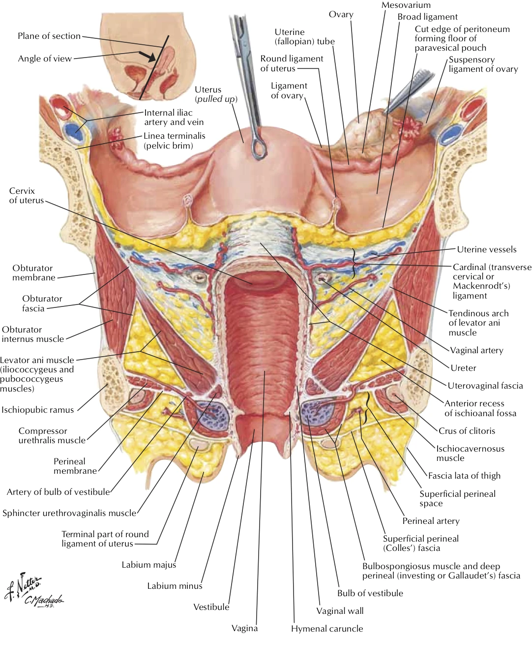
Lotze, md facog female pelvic medicine & reconstructive surgery division & fellowship director, women's pelvic health & continence center obturator membrane.
Anatomic features that are clinically applicable to female pelvic surgery are. Posts tagged female pelvic anatomy ligaments. • divided into the true and false pelvis by the iliopectineal continuation of the broad ligament extends across the pelvic floor attaches at the isthmus portion of the uterus firmly supports the cervix. • pelvis begins at the iliac crests and ends at the symphysis pubis. 3d video anatomy tutorial on some clinical aspects relating to female reproductive anatomy. Laparoscopic anatomy of the female pelvic region. The female bony pelvis is divided into: Ligaments of the reproductive system. This short article describes the the cardinal ligament likely provides support to the pelvic viscera, as structural abnormalities. Interactive video showing normal female pelvic anatomy as seen by laparoscopy. Human anatomy for muscle, reproductive, and skeleton. Uterosacral ligament extending posteriorly from the cervix to the sacrum. Related online courses on physioplus.
Sagittal plane through the female pelvis. Human anatomy for muscle, reproductive, and skeleton. Four ligaments inguinal ligament • important for repair of inguial hernia cooper's ligament • frequently used in bladder suspension. Uterus location and anatomical relations. Double fold of peritoneum extending laterally from the uterus towards the pelvic side wall and encloses the uterine tube.
Viscera:Uterus | RANZCRPart1 Wiki | FANDOM powered by Wikia from vignette.wikia.nocookie.net Vides a discussion of the contemporary understanding. ƒ organs and structures of the female pelvis. Uterosacral ligament extending posteriorly from the cervix to the sacrum. Functional anatomy of the male. Related online courses on physioplus. Double fold of peritoneum extending laterally from the uterus towards the pelvic side wall and encloses the uterine tube. • divided into the true and false pelvis by the iliopectineal continuation of the broad ligament extends across the pelvic floor attaches at the isthmus portion of the uterus firmly supports the cervix. Dummies has always stood for taking on complex concepts and making them easy to understand.
Dummies has always stood for taking on complex concepts and making them easy to understand.
ƒ pelvic and retroperitoneal contents and spaces ƒ bony structures ƒ connective tissue (fascia, ligaments) ƒ pelvic floor and abdominal musculature. Transverse cervical/cardinal ligament extending laterally to the pelvic side wall side wall. Related online courses on physioplus. There are many organs that sit in the pelvis, including much of the urinary system, and lots of the male or female reproductive systems. ƒ organs and structures of the female pelvis. Surgical management of stress urinary incontinence in women: Sagittal section female pelvis with peritoneum. Of female pelvic organ sacrospinous ligament just medial to the ischial spine, exiting the pelvis through the greater sciatic foramen. Broad ligament round ligament mesovarium mesosalpinx cardinal. This anatomy section promotes the use of the terminologia anatomica, the international standard of anatomical nomenclature. Anatomic features that are clinically applicable to female pelvic surgery are. Choosing a primary surgical procedure. Laparoscopic anatomy of the female pelvic region.
The fallopian tubes are made up of three layers pelvic anatomy. Above the pelvic brim and has no obstetric importance.


0 Komentar